🎯 Learning Outcomes
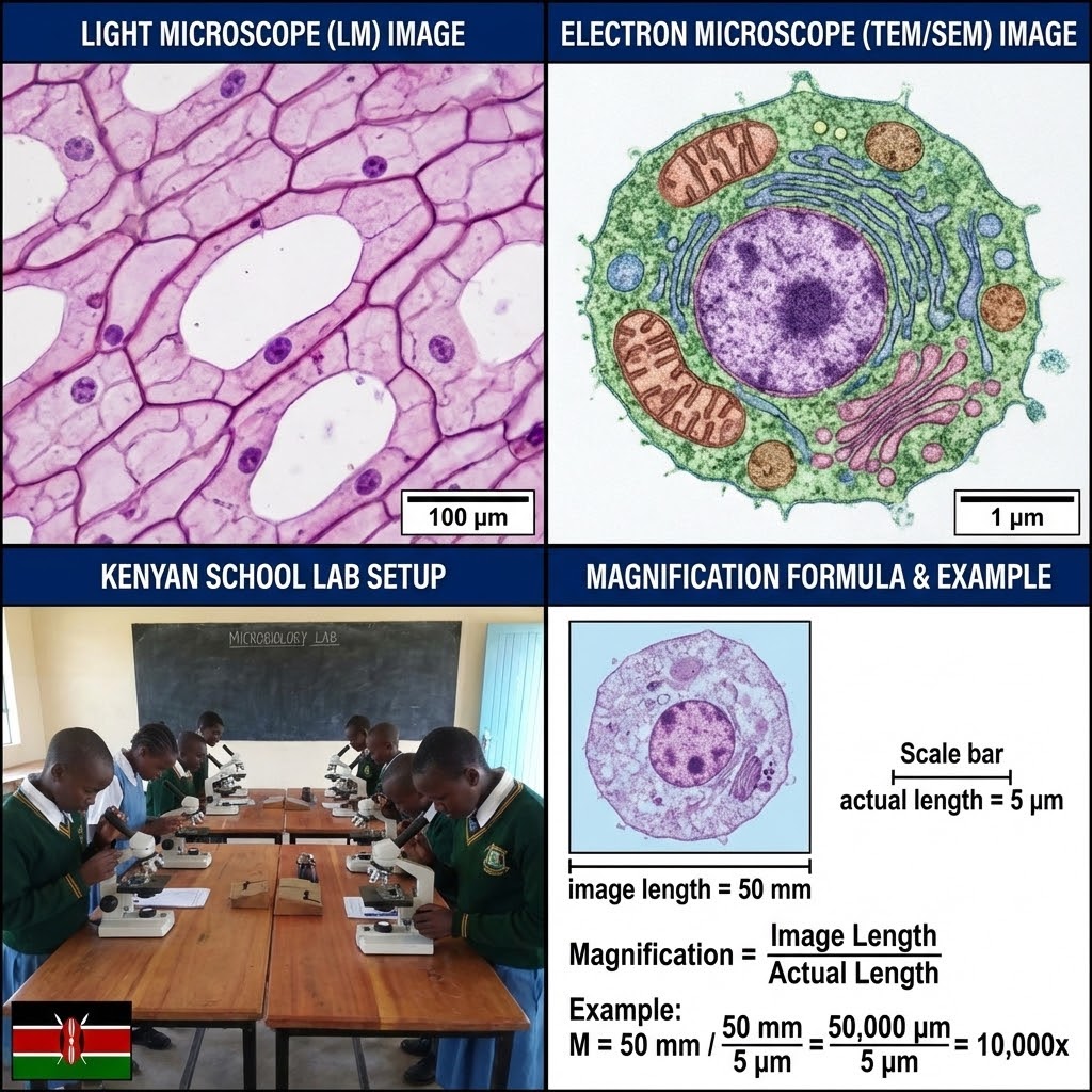
- Distinguish between Light and Electron microscopy resolutions.
- Execute technical procedures for temporary slide preparation.
- Describe cellular organelles and their ultrastructural functions.
- Calculate cell size using the magnification formula.
- Explain how specialized cells are structurally adapted for physiological roles.
🔬 Microscopy & Magnification
In Biology, we must calculate the true size of organisms. Because images are magnified, we use a specific ratio to find the actual size.
If a student observes a cell under a microscope and the drawing measures 40mm, but the actual cell is only 0.02mm, the magnification is:
40mm / 0.02mm = 2000x
| Feature | Light Microscope (LM) | Electron Microscope (EM) |
|---|---|---|
| Resolution | Low (200nm) - Cannot see ribosomes | High (0.5nm) - Sees detailed ultrastructure |
| Specimen Prep | Simple (Sectioning & Staining) | Complex (Dehydration, Vacuum, Gold coating) |
Grade 10 CBE Kenyan Examples & Practical Notes:
- Light microscope: Used in Kenyan schools — max ~1500×, good for whole cells (e.g., onion epidermis, cheek cells), but cannot see organelles like mitochondria clearly.
- Electron microscope: Used in university/research labs (e.g., University of Nairobi, KEMRI) — shows ultrastructure (ribosomes, cristae in mitochondria).
- Magnification calculation: Common exam question — always convert units (mm to μm: 1 mm = 1000 μm) before dividing.
- Lab activity: Prepare onion epidermis slide → measure cell length on drawing → calculate actual size using given magnification → compare with textbook value.
- Misconception to correct: "Higher magnification = better view" — no, resolution (ability to separate close points) matters more than magnification; beyond ~1500× light microscope resolution is lost.
🧫 Technical Slide Preparation
To view cells clearly, we follow a rigorous preparation protocol. Senior learners must master these four techniques:
Grade 10 CBE Kenyan Lab Tips & Safety:
- Common slides: Onion epidermis (easy, shows cell wall, nucleus), cheek cells (human epithelial), Elodea leaf (shows chloroplasts, streaming cytoplasm).
- Stains: Iodine (starch — blue-black), methylene blue (nucleus — blue), safranin (lignin in xylem — red).
- Safety: Wear gloves when handling stains, avoid eye contact, dispose slides properly (not in sink), clean microscopes after use.
- Lab activity: Prepare temporary mounts of onion epidermis → stain with iodine → observe under low/high power → draw and label (cell wall, cytoplasm, nucleus).
- Misconception to correct: "All slides need permanent mounting" — no, temporary mounts (water/glycerine) are quick and sufficient for school practicals.
🏗️ Organelle Ultrastructure
As observed under the Electron Microscope, cells contain highly specialized compartments:
| Organelle | Ultrastructural Description | Function |
|---|---|---|
| Mitochondria | Double-membraned; inner membrane folded into cristae. | Primary site of ATP (energy) production via aerobic respiration. |
| Ribosomes | Small dense granules; can be free-floating or on Rough ER. | The site of protein synthesis (translation). |
| Chloroplast | Contains stacks of thylakoids (grana) in a fluid stroma. | Site of photosynthesis; contains chlorophyll to trap light. |
| Nucleus | Double-membraned with nuclear pores and a nucleolus. | Stores genetic information; controls all cellular activities. |
Grade 10 CBE Kenyan Examples & Functions:
- Mitochondria: Abundant in muscle cells (e.g., runner's legs), flight muscles of birds — "powerhouses" for ATP.
- Ribosomes: Free in cytoplasm (make proteins for cell use) or on rough ER (make secreted proteins, e.g., enzymes in salivary glands).
- Chloroplasts: In palisade mesophyll of maize/sugarcane leaves — site of photosynthesis (food production for Kenyan staple crops).
- Nucleus: Contains chromosomes (DNA) — controls cell division, protein synthesis; nucleolus makes ribosomes.
- Misconception to correct: "All cells have chloroplasts" — no, only plant cells (and some protists); animal cells do not photosynthesize.
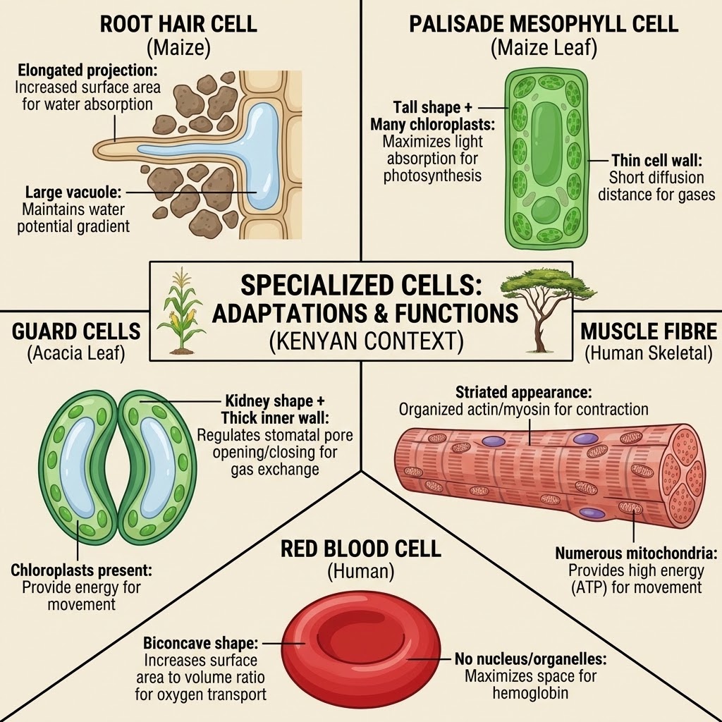
🧬 Cell Specialization & Adaptation
Cells do not all look the same. They undergo differentiation to become efficient at specific tasks.
1. Root Hair Cell (Plant): Has a long, thin extension. This increases the surface area to volume ratio, allowing for faster absorption of water and mineral salts from the soil.
2. Muscle Cell (Animal): Contains a very high density of mitochondria and contractile filaments. This allows the cell to produce the massive amounts of energy required for contraction and movement.
3. Guard Cells (Plant): Found in pairs around the stoma. Their cell walls are thicker on the inner side, allowing them to curve and open the pore when turgid.
Grade 10 CBE Kenyan Examples & Adaptations:
- Root hair cells: In maize, beans — long projection increases surface area for water/mineral uptake from Kenyan soils (often nutrient-poor).
- Muscle cells: Skeletal muscle (biceps in humans) — many mitochondria for ATP, myofibrils for contraction; cardiac muscle — branched, intercalated discs for synchronized beating.
- Guard cells: In maize/sorghum leaves — kidney-shaped, thick inner wall → open/close stomata to control water loss in dry Kenyan climate.
- Other examples: Palisade mesophyll (tall, packed chloroplasts for max photosynthesis), xylem vessel elements (dead, hollow tubes for water transport), red blood cells (biconcave, no nucleus for max oxygen carry).
- Lab activity: Observe prepared slides (root hair, muscle, leaf epidermis) → draw and label adaptations → link structure to function.
- Misconception to correct: "All cells are the same size/shape" — no, specialization (differentiation) leads to huge variation in form and function.
❓ Inquiry Question
Answer: Cells are specialized through structural modifications. By changing their shape (e.g., biconcave in red blood cells) or increasing the number of certain organelles (e.g., many chloroplasts in palisade cells), they become optimized for their specific biological role.